Lung Functions and Control of Breathing
| ✓ Paper Type: Free Assignment | ✓ Study Level: University / Undergraduate |
| ✓ Wordcount: 1386 words | ✓ Published: 12 Oct 2017 |
Lung Functions and Control of Breathing
ABSTRACT
The importance of oxygen for human body can neither be under nor over estimated and this “essential for life” function of oxygen delivery to body cells is performed by the RESPIRATORY SYSTEM through the process of BREATHING. Not only this, breathing helps body get rid of the waste product of cellular respiration, i.e. carbon dioxide. This exchange of oxygen and carbon dioxide occurs at the level of lungs in the respiratory system. Even though breathing is an unconscious affair, something we are not even aware of doing all the time, our body has many ways of regulating and controlling this function through a complex mechanism of interdependent pathways.
RESPIRATORY SYSTEM
Definition
Respiratory system is an integrated system of organs involved in the intake and exchange of oxygen and carbon dioxide between an organism and the environment.
Components
The respiratory system is made up of:
- Nose
- Pharynx
- Larynx
- Trachea
- Bronchi
- Lungs
The primary function of the respiratory system is to supply the blood with oxygen in order for the blood to deliver oxygen to all parts of the body and to remove carbon dioxide.
This process starts with the oxygen rich air entering the nose or mouth which then moves down the trachea, bronchi and finally reaches the lungs.
Each of the organs of respiratory system thus plays an important part in the process of respiration but the lungs are the site where actual exchange of gases takes place.
LUNGS
The lungs are a pair of cone-shaped, soft, spongy, pinkish organs. They are located in the anterior chest or the thorax.
Functions of Lungs
Two types of functions are performed by lungs:
- Respiratory functions
- Non-respiratory functions
Respitory Functions
The primary function of lungs is “gas exchange” i.e. to exchange oxygen from air with carbon dioxide from blood. The process in which this happens is called “external respiration” or breathing.
The average adult’s lungs contain about 600 million spongy, air-filled sacs called alveoli that are surrounded by capillaries. The inhaled oxygen passes into the alveoli and then diffuses through the capillaries into the arterial blood. Meanwhile, the waste-rich blood from the veins releases its carbon dioxide into the alveoli. The carbon dioxide then follows the same path out of the lungs (as oxygen coming inside the lungs) when we exhale.
Non-Respitory Functions
These are categorized as:
- Mechanical
- Biochemical
- Physiological
The lungs defend the body from potentially dangerous airborne pollutants and toxic chemicals produced by our own body. They are the first line of defense against airborne bacterial and viral agents, other infectious agents and irritants. The lungs control the flow of water, ions and proteins across its cellular structures. The lungs manufacture certain essential hormones and other chemicals for very specific functions in the body. They remove volatile particles and substances generated within the body. With the liver, they act as the metabolic product “removals services” of the body.

CONTROL OF BREATHING
Main goals of the respiratory control system
- Rate of Alveolar ventilation sufficient to maintain the normal level of blood gases.
- Changes in alveolar ventilation rate sufficient to acclimatize changing environments or metabolic needs (e.g. jogging).
- Adaptability to allow other activities such as talking or eating which share anatomical structures with the lung.
Two systems control breathing
- Involuntary Control
- Voluntary Control
Involuntary Control
Involuntary Control is mediated by the brain’s respiratory center located in the brainstem, particularly in the medulla oblongata (has the inspiratory centre),pons (has the expiratory centre). The inspiratory center sets the basic rhythm by automatically initiating inspiration with a two second burst of nerve impulses to the diaphragm and the external intercostal muscles.Contraction of the diaphragmand the external intercostal muscles draws air into the lungs. The neurons stop firing for about three seconds and the muscles relax.The elastic recoil of the lungs and chest wall results in expiration. The expiratory center functions during forced expiration stimulating the internal intercostal and abdominal muscles to contract.
Voluntary Control
The normal involuntary nature of the respiratory cycle can be suppressed or altered to meet the changing metabolic demands of the body.Neural and chemical factors regulate both the rhythm and the rate and depth of ventilation.
Respiratory control system involving the nervous system has three components:
Sensors and their afferents
Providing information on what the system is doing.
Central Controller
Compares intended operation with how the sensors say the system is actually working.
Efferents
These are respiratory muscles which actually carry out respiration.
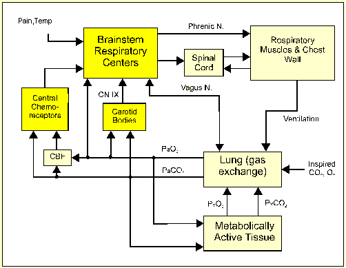
Block diagram showing major components of ventilatory control.
Central Controlling Area
The central controlling area for breathing, called the respiratory centre, is located in the brainstem. There are “inspiratory neurons” which are active during inspiration and inactive during expiration. Other neurons are active during expiration but not inspiration-the “expiratory neurons”. These two groups of neurons automatically maintain a rhythmic cycling pattern of inspiration and expiration. This automatic rhythm can be modified by the afferent information.
Afferent Supply
- Central chemoreceptor
Chemo receptors are cells that respond to chemical stimuli. There are cells in the floor of the fourth ventricle and are responsive to arterial Pco2 by way of hydrogen ion concentration in cerebrospinal fluid (CSF).Carbon dioxide in the blood can rapidly diffuse across into the CSF, and there is a balance between the level of carbon dioxide, hydrogen ion and bicarbonate ion in the CSF. If the carbon dioxide in the blood increases (e.g. following exercise), then the carbon dioxide, hydrogen ion and bicarbonate ion concentrations increase correspondingly in the CSF. This increase in CSF acidity causes hyperventilation which lowers the carbon dioxide concentration in the blood. A low blood carbon dioxide level has the opposite effect and leads to hypoventilation.
- Peripheral chemoreceptor
These are small pieces of tissue that contain chemoreceptors which respond to the oxygen and carbon dioxide concentrations in arterial blood.
Carotid bodies located at bifurcation of common carotid. Carotid bodies are afferent in glossopharyngeal nerve.
The output from the carotid body is thought to provide information to allow immediate regulation of breathing, breath by breath, by the respiratory centre. In normal people, if the arterial blood reaching the carotid body has a partial pressure of oxygen of 10kPa (80mmHg) or a carbon dioxide partial pressure of more than approximately 5kPa, (40mmHg), then there is an immediate and marked increase in breathing.
Aortic bodies found between ascending aorta and pulmonary artery. Aortic bodies are afferent in vagus nerve.
The aortic bodies seem to respond somewhat to changes in oxygen content such as are seen in anemia. This is due to the fact that the arterial-venous oxygen difference is greater for these cells than for the carotid bodies. Thus, the receptor cells see lower average oxygen when content is reduced.
- Brain
Breathing can be influenced by other parts of the brain. We can all consciously breathe deeply and more rapidly (called hyperventilation), and this can happen, for example, before starting strenuous exercise, Intensely emotional situations, part of the response to massive blood loss. This response is coordinated by the autonomic system in the hypothalamus and the vasomotor centre in the brain stem.
- Lung
There are various receptors in the lung that modify breathing. Receptors in the wall of the bronchi respond to irritant substances. In the elastic tissues of the lung and the chest wall are receptors that respond to stretch. The stretch responses occur when the lung and chest wall are distended and inhibit further inspiration. Conversely, when the lung volume is low, then there are opposite reflexes.
Efferent
The efferent nerves from the respiratory centre pass down the spinal cord to the diaphragm, intercostal muscles and accessory muscles of inspiration in the neck. During normal breathing, inspiration is an active muscular process. Expiration is passive and relies on the natural elasticity of the tissues to deflate the lung.
REFERENCES
Text book of Medical Physiology, 9th Edition, Guyton & Hall, Saunders
The Lungs in Health and Disease – Pamphlet, National Heart, Lung, and Blood Institute
Introductory Anatomy: Respiratory System, .R.Johnson
Review of Medical Physiology, William Ganong
Anatomy & Physiology, McGraw-Hill
Clinical Respiratory Physiology, Taylor, AE, K Rehden, RE Hyatt, and JC Parker
Neuronal Connections of a Ventral Brainstem Respiratory Chemosensitive Area, Paul, Anthony D
Breathing Coordination, Rabbany, Sina Y
Pulmonary Curriculum Function: Neural Control of Breathing, Webber, Charles L., Jr.
Human Physiology 4th ed., Rhoades, Rodney, Pflanzer, Richard
Cite This Work
To export a reference to this article please select a referencing stye below:
Related Services
View all


DMCA / Removal Request
If you are the original writer of this assignment and no longer wish to have your work published on UKEssays.com then please click the following link to email our support team:
Request essay removal